Real-Time Ultrasound Core Training
Ultrasound Core
Core stability is the power of the trunk to control movement and reduce the painful stress on the lumbar and pelvic spine. The spine is usually unstable and depends on ligaments and muscles surrounding it for stability. Arm and leg movements like walking, running, and picking things up are absorbed, redirected, and transferred through the lumbopelvic spine.
Two types of muscles are associated with core stability movers and stabilisers. Large, powerful muscles are responsible for movement, and locomotion comes under movers. Stabilisers have smaller and deeper muscles that protect and stabilise the lumbopelvic spine by creating low force and avoiding abnormal vertebral movements that can cause severe injuries.
The deep stabilising muscles of the lumbopelvic spine, which are attached to the lumbar spine and pelvis, give the required support and control to the spine. This stabilisation assists larger mover muscles in functioning correctly, especially when lifting, moving heavy objects, and doing sports activities. Research indicates that core stability needs properly coordinated activation of these deep stabilising muscles before movement occurs. If you have a back injury, these stabilising muscles can become numb and stiff. It may remain the same if it is not treated correctly as it is sensitive.
What are the Primary Core Muscles?
- Multifidus
- A small muscle that connects each vertebra and the pelvis. Its close positioning to the spine’s rotation centre helps it stabilise the lumbar spine by restricting abnormal movements and increasing spinal fitness.
- Transverse Abdominis
- This is the deepest layer of the abdominal muscles, forming a thin sheet that connects to the spine. When activated, it braces the abdomen and stabilises the lumbar pelvic region.
- Diaphragm
- A dome-shaped muscle separates the lungs from the abdomen, controlling breathing and the spine. When you inhale, it increases the intra-abdominal pressure, improving the stabilising effect of the transverse abdominis.
- Pelvic Floor Muscles
- These muscles form a diaphragm that supports the abdominal organs, including the bladder, intestines and uterus. They keep the intra-abdominal pressure short, called IAP, in position, which is essential for spinal stability. Poor pelvic floor activation can increase back pain and urinary stress incontinence, which affects women post-childbirth, aged people, and those who are recovering from complicated surgeries like hysterectomy or prostrate operations.
What are the Primary Core Muscles?
Core training benefits individuals suffering from back pain, sacroiliac joint instability, pelvic floor dysfunctions, sciatica, and shoulder or hip dysfunctions. These conditions get aggravated if core stability weakens. Focusing on strengthening these deep stabilisers eliminates symptoms and improves body functioning.
Benefits of Real-Time Ultrasound Core Training
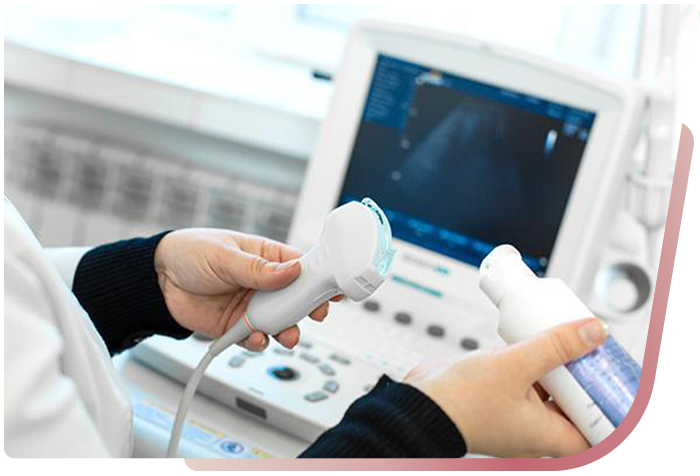
Core muscles are deep and generate low force, making it challenging for patients and doctors to diagnose accurately. Real-time ultrasound offers a non-invasive solution, allowing doctors and patients to visualise muscle activation. This helps activate and engage the core muscles during the training process.
Real-time ultrasound core training focuses on shifting these muscles in a coordinated and safe way without painful force. This method is valuable for those with pain from injury as it helps recover the muscles effectively. RTUS core training has six sessions combined with a home exercise program, which sets you off to regular exercises and enables you to avoid injury quickly.
Our team of professional doctors offers proper feedback and guides you through a healthier and happier lifestyle. Contact our team and become the most active version of yourself!